Left abomasal displacement (LDA) is a common and economically important disease in high-yielding dairy cows. Since the first description of LDA in 1950, many techniques for surgical correction of this disorder have been developed. At Giessen University in Germany, the standard procedure to correct LDAs is the minimally invasive repositioning of the displaced abomasum according to the Janowitz method, first described in 1998.

In the last 11 years, 2,450 cows suffering from LDA underwent surgery at our clinic. More than 420 were treated by the Dirksen method and 1,958 according to the Janowitz method.
Since the introduction of the minimally invasive repositioning of left displaced abomasum, known as the Janowitz method, this technique has become the most commonly used procedure for the correction of this disorder at our clinic.

Besides this laparoscopic method, the left or right flank laparotomy with omentopexy (Utrecht or Dirksen method) are routinely used in pregnant cows or in patients that suffer from a thrombophlebitis of the udder veins or an oedema of the udder itself.
The two-step Janowitz method to correct a displaced abomasum has been modified into a one-step method. Since the year 2000, this technique is shown besides the classical Janowitz operation at the International Workshop on Laparoscopic Diagnostics and Therapy in cattle, which takes place once a year at our clinic.
Materials and methods
Research showed a comparison of success rates of laparoscopy-assisted abomasopexy versus omentopexy via right flank laparotomy for the treatment of dairy cows with left displaced abomasum (LDA). They could show success rates for both methods were almost equal.

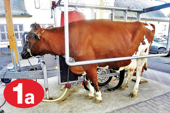
The laparoscopic operation, according to Janowitz, is basically performed as follows: After the clinical examination of the cow, she is prepared for endoscopic surgery by clipping and aseptically preparing two small areas at the left side of the abdominal wall of the cow ( Figures 1a+1b ).
Position 1 is meant for the endoscope in the left paralumbar fossa; position 2, for the toggle setting trocar, is situated between the last two ribs. Local anaesthesia is performed in both positions, whereas sedation of cows is necessary in very nervous cases only.
With the introduced endoscope, the left displaced abomasum can be seen as a dome between the left abdominal wall and the rumen on the right side. Cranially, the spleen and the diaphragm can be seen.

Under visual control, the toggle pin can be put into the abomasum on top of the organ via a toggle setting trocar ( Figure 2a ). Through this trocar, deflation of the abomasum is performed ( Figure 2b ).
After complete deflation of the abomasum, part two of the operation is prepared, i.e. the patient is brought into dorsal recumbency. Front and hind legs have to be tied to guarantee a safe performance of surgery.
On the ventral abdominal wall, two more small areas are prepared aseptically for surgery. Again, one is meant for the optics and the other one is the point of fixation for the formerly displaced abomasums ( Figure 3 ).

Fixation of the abomasum is performed in right lateral recumbency by gentle tension to the sutures of the toggle pin ( Figure 4a ) and knotting them to a gauze bandage ( Figure 4b ).
Insertion sights of the trocars (both, ventrally and on the left side) are covered with aluminium spray and the patient is brought into an upright position.
The gauze bandage is left in place for three to four weeks before removal by the owner. According to the original description of this method, antibiotics or analgetics that would imply withdrawal times are not necessary after routine surgery.

Conclusions
Within the last 10 years, the Janowitz method has become the standard surgical procedure for the correction of left displaced abomasums at our clinic.
Complications due to dorsal recumbency are seen very seldom and, subject to the condition that surgery is performed very carefully, the advantages of the procedure exceed its disadvantages by far. PD
References omitted due to space but available upon request. Click here to email an editor.
Sickinger is a researcher at the Clinic for Ruminants (Internal Medicine and Surgery) at Justus-Liebig-University in Giessen, Germany.
PHOTOS:
TOP RIGHT: Figures 1a+1b – Preparation of the patient for endoscopic surgery
TOP MIDDLE RIGHT: Figure 2a – Insertion of the toggle setting trocar into the displaced abomasum
MIDDLE RIGHT: Figure 2b – Deflation of the abomasum and insertion of the toggle sutures into the abdominal cavity
MIDDLE RIGHT: Figure 3 – Grasping of the toggle sutures
BOTTOM MIDDLE RIGHT: Figure 4a – Toggle sutures for fixation of the abomasum
BOTTOM RIGHT: Figure 4b – Ventral fixation of the abomasum with gauze bandage.


