“Approximately 70 percent of all eye tumors in cattle are cancerous,” says Jeremy Powell, DVM, University of Arkansas. “Cancer eye is an aggressive disease that can infiltrate tissue surrounding the affected eye and invade lymph nodes near the lesion.

Tumors that result in extensive invasion can impair a cow’s ability to raise her next calf. Once a cow is noted to have the disease, a decision should be made to cull or treat.”
“When a tumor has spread to the extent that it has destroyed the eye, the animal is condemned at the processing plant during inspection before slaughter,” says Dr. William James, the USDA’s former chief veterinarian.
“If the tumor is relatively small, the animal is tagged ‘suspect’ and is slaughtered, and the public health veterinarian (PHV) inspects it at post-mortem. Significant spread of the tumor around the eye, which is visible after hide removal, or development of secondary malignant growths in the lymph nodes of the head causes the entire animal to be condemned.
If the tumor is localized, the head is condemned and the carcass and internal organs passed, assuming no other condemnable diseases or conditions are found.”
Detection and therapy
“Many producers are dismayed to find animals afflicted with cancer eye that were clean a few months previously,” states Larry Foster, Ph.D., extension beef cattle specialist, New Mexico State University. “This unpleasant experience can be avoided by learning to detect early eye tumors, which are not yet cancerous, and treating them before they turn malignant. Producers can practice preventive medicine in the case of cancer eye.
“Most people have no trouble recognizing cancer eye, yet few recognize benign or precursor lesions, which are highly treatable. Precursor lesions on the eyeball are known as plaques or papillomas.
They are easily recognized as white or pink growths at the edge of the colored part of the eye (corneoscleral region). Lesions in the center of the pupil are usually the result of pinkeye or physical damage and are usually not precancerous.”
The third eyelid is a common site for malignant tumors. It is transparent or translucent and can be drawn across the eye for protection and provide moisture while maintaining visibility.
On the upper and lower eyelids, the precursor lesions are known as keratomas, commonly called “wickers.” Usually occurring on the lower lid, these small tumors are often crusted over with scab-like material. If the growth appears to be attached to the eyelash, it is probably dried eye matter.
When the growth appears to be attached directly to the lid, and removal of the scab reveals a small growth and perhaps a bit of bleeding, it is probably a precursor lesion and is highly treatable.
“Probably the easiest and most effective treatment of these benign lesions is a localized current field (LCF) radio frequency energy to heat the tumor,” says Mel Pence, DVM, University of Georgia. “Since cancer cells have a large nucleus, they are more susceptible to heat than healthy cells with a small nucleus.
This method of treatment heats the cells to the point that the cancer is destroyed while leaving healthy cells intact. Cryosurgery (freezing with liquid nitrogen) can be successful on small tumors, but it also leaves a scar on the eye surface.
“There are two important considerations about cancer eye treatment: No method is 100 percent sure, and all treatment should be considered a temporary procedure,” Pence continues. “For example, you may treat a cow with a small calf at her side to allow her to raise the calf – and then sell both in the fall.
Experience indicates that once a cow has cancer eye, she will probably get it again, although it usually will occur somewhere else or in the other eye.”
Cancer eye rarely occurs on dark-colored eyelids. Pigmented eyelids are moderately heritable with a heritability estimate of about 0.4. This means that 40 percent of the tendency for a calf to have pigmented eyelids comes from its parents’ genetics. The heritability estimate for pigmented eyelids is similar to that of birthweight.
Eye pigmentation measurement
A modern protocol for assessing eye pigmentation was developed by Kaycee Davis, a graduate student working under the direction of David Riley, Ph.D., Texas A&M University. Davis assessed variation in eyelid and corneoscleral pigmentation by counting pixels in images and calculating percentages of pigmented areas.
We may need to re-study parts of the eye in order to understand cancer eye and Davis’ study. The pupil is the opening in the center of the iris, which is a thin, circular structure. It is responsible for controlling diameter and size of the pupil and giving the eye its color. A transparent layer above the iris and pupil is called the cornea. The sclera encircles the iris and is the white of the eye.
“Pale pink is considered to indicate absence of pigment,” Davis says. “Corneoscleral pigment is not fully developed at birth but increases after 5 years old until about 9 years old. Amounts of pigmentation on the corneoscleral region are related to the quantity on eyelids. The non-haired region is the eyelid target for tumors.”
A total of 1,083 head of purebred Herefords and Hereford crosses with Bos taurus and Bos indicus were evaluated. The cattle were on farms and ranches in Texas, Oklahoma, Arkansas, Louisiana, Mississippi, Florida, North Carolina and South Carolina. Age categories of the cattle were calf, yearling and mature. Both female and male animals were evaluated.
Two photographs were taken of each eye while the animal was restrained in a squeeze chute. Davis sorted the photographs into similar data groups and discarded those that could not be used due to poor quality.
The usable photographs were cropped in Adobe Photoshop Elements 2.0 to provide images of the eye parts of concern. Final count of test samples was 564 eyelids and 1,033 corneoscleral regions that were partially pigmented. Fully pigmented eyes and eyes without any pigment were not included in these totals.
Davis was able to quantify the amount of pigment in each sample through digital analysis. Her study results were consistent with information obtained from a series of research investigations done in the 1950s and 1960s.
She found a lower incidence of precancerous to cancerous lesions in white-face cattle with increased pigment around the eyes. Mature animals had more corneoscleral pigmentation than did young animals. The amount of pigmentation varied among breed types. There was no difference in pigmentation between sexes, between calves and yearlings, and between the right and left eye.
The study, completed by Davis in 2013, provides a better understanding of the relation of eye pigmentation to cancer eye. ![]()
Robert Fears is a freelance writer based in Texas.
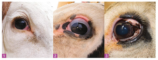
PHOTOS
1: The upper eyelid is pigmented on this animal and the lower eyelid does not have pigment.
2: In this eye, there is no presence of lesions, zero corneoscleral pigmentation, 45.5 percent upper eyelid pigmentation and 70.2 percent lower eyelid pigmentation.
3: This eye has no presence of lesions, 42.7 percent corneoscleral pigmentation, 40.7 percent upper eyelid pigmentation and 54.1 percent lower eyelid pigmentation. Photos courtesy of Kaycee Davis, graduate studies, animal breeding and genetics, Texas A&M University.
Management practices to control cancer eye in beef cattle
- Early detection is a key control factor. Check eyes for lesions when cattle are put through the chute for other reasons.
- Consult with your veterinarian on whether precancerous lesions and tumors should be removed.
- Take bulls out of service if they have a tendency for cancer eye so they won’t pass the trait to their offspring.
- When buying herd replacements, select animals with pigment around their eyes.
- Consider keeping cows with cancer eye as long as they produce a good healthy calf every year.
- When animals with cancer eye are to be sold for slaughter, try to do so before the cancer spreads in order to avoid
carcass condemnation.


